In short
A lung nodule is a small round spot seen on CT. More than half of single nodules are not cancer, and size and whether it is solid or ground-glass decide follow-up. In low-risk adults aged 35 and over, the Fleischner guideline needs no routine follow-up under 6 mm; larger ones get repeat CT scans, PET/CT or a biopsy.
Emergency if: You are coughing up more than a few spots or streaks of blood, or you cough up blood and find it hard to breathe, have a very fast heartbeat or have chest pain; or you are struggling to breathe, gasping and unable to speak; or you have pain in your chest or upper back: call emergency services now or go to an emergency department now. The NHS lists these for immediate action on its coughing up blood and lung cancer pages. Do not drive yourself.
On this page
01
What is a lung nodule on a CT report, and is it cancer?
MedlinePlus describes a pulmonary nodule as a round or oval spot in the lung seen on a chest x-ray or CT scan. RadiologyInfo says a lung nodule is a small growth, under 3 cm, that is common: nodules are found in up to half of adults who have chest x-rays or CT scans, usually on a scan done for another reason, and a person may have one or several. A report line such as nodule in the right upper lobe is the radiologist describing that spot.
More than half are not cancer. MedlinePlus says more than half of solitary pulmonary nodules are noncancerous and that scars and past infections are common causes: infectious granulomas, formed as a reaction to a past infection such as tuberculosis or a fungal infection, cause most benign nodules. RadiologyInfo says small lung nodules are typically benign, though some may be early-stage lung cancer, and that most cause no symptoms. A nodule that is not cancer can still need a repeat scan to be sure, which is why follow-up exists.
02
What size of lung nodule is a worry? What do 4 mm, 6 mm and 8 mm mean?
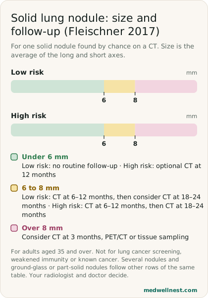
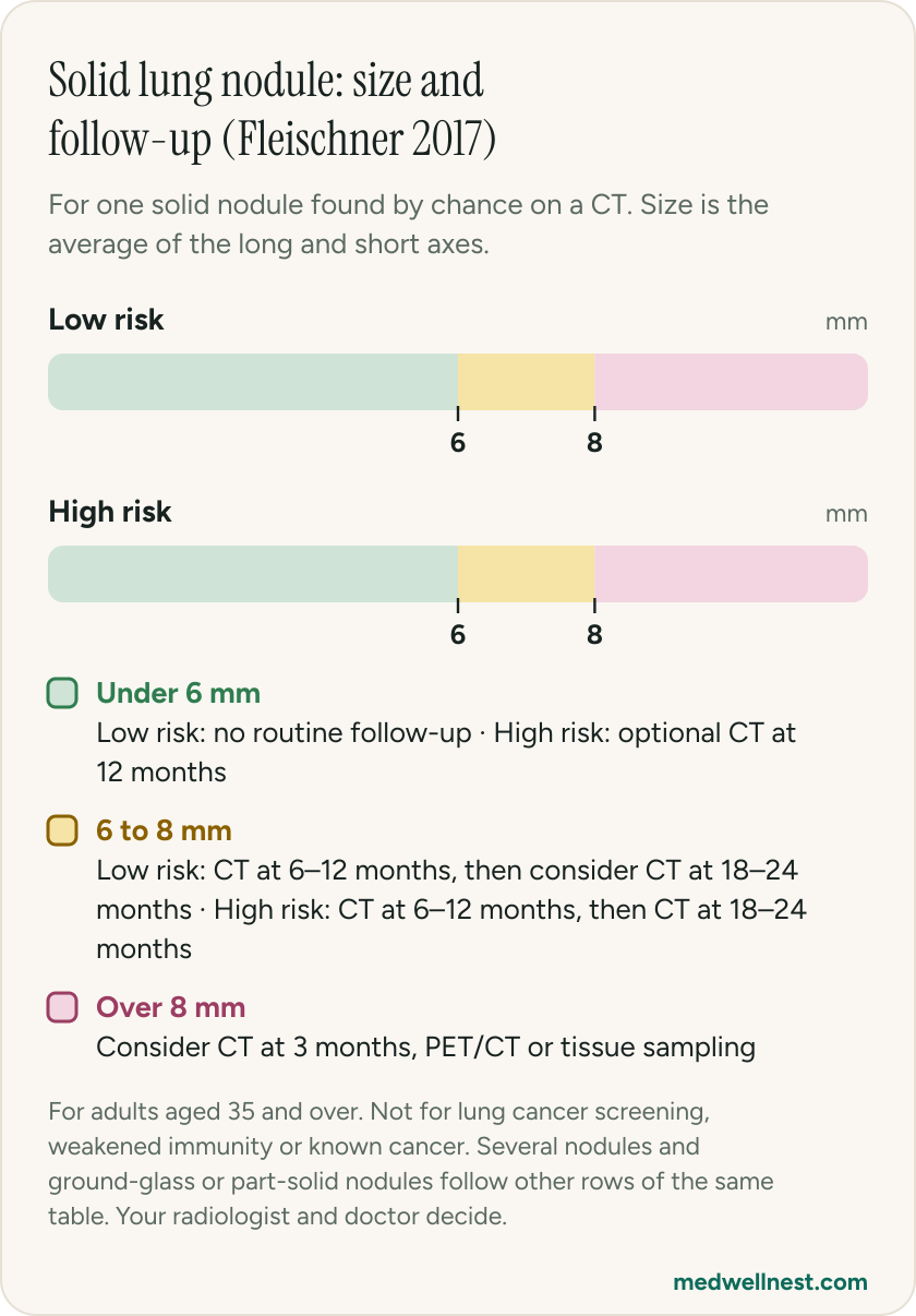
The Fleischner Society 2017 guideline, from the journal Radiology, raised the minimum size for routine follow-up and gives recommended intervals as a range. A 2026 review reproducing its table says the 6 mm threshold for a solid nodule is based on a cancer risk estimate of 1% or more, and that nodules under 6 mm generally need no follow-up in low-risk people unless they have high-risk features such as spiculated or irregular margins or an upper lobe position, in which case a CT at 12 months may be needed.
In plain words, for one solid nodule: under 6 mm, no routine follow-up in a low-risk person and an optional CT at 12 months in a high-risk person. From 6 to 8 mm, a CT at 6 to 12 months, then consider a CT at 18 to 24 months for low risk, or a CT at 18 to 24 months for high risk. Over 8 mm, consider a CT at 3 months, a PET/CT scan or a tissue sample. Your doctor judges the risk group from things such as smoking history, age, family history and the nodule's size, edge and position (see below). Size means the average of the long and short axes rounded to the nearest millimetre, so a report that gives one longest dimension can be slightly bigger than the guideline size. Ask your radiologist which was used.
03
What is a ground-glass or part-solid nodule? Is it different?
Nodules are described by how dense they look. The 2026 review says a solid nodule has soft-tissue density, while a subsolid nodule is either ground-glass, a hazy look, or part-solid, with both hazy and solid parts. Part-solid nodules have been linked with a higher risk of lung cancer than solid ones, and pure ground-glass nodules with a lower risk than solid ones. Subsolid nodules also grow slowly and may need follow-up for longer.
For a single ground-glass nodule under 6 mm the Fleischner table says no routine follow-up, and from 6 mm it says a CT at 6 to 12 months to check it persists, then a CT every 2 years until 5 years. For a single part-solid nodule under 6 mm it says no routine follow-up, and from 6 mm it says a CT at 3 to 6 months, then yearly CT for 5 years if it is unchanged and the solid part stays under 6 mm. A persistent part-solid nodule with a solid part of 6 mm or more is called highly suspicious and is looked at more closely.
04
What else besides size changes the plan? Does smoking matter?
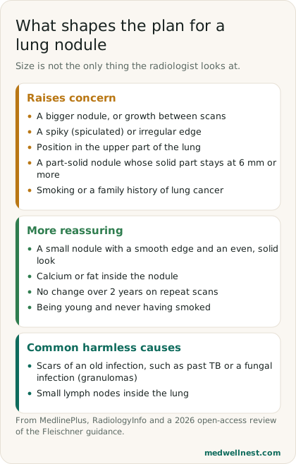
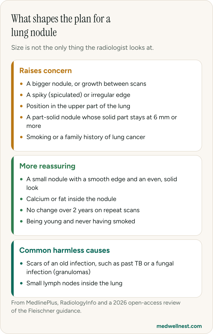
RadiologyInfo lists what raises the chance that a nodule is cancer: being over 50, smoking, a family history of lung cancer, symptoms of lung cancer, a larger size, growth over time and a spiky (spiculated) shape. The 2026 review adds that lung cancer favours the upper lobes, that calcium or fat inside a nodule can suggest a benign nodule, and that a smoker has a 10 to 35 times higher risk of lung cancer than a non-smoker. Our stories on quitting smoking and on lung cancer cover those topics.
MedlinePlus says a nodule is more likely benign if it is small, has a smooth border and a solid, even look, and you are young and have never smoked. It says that if repeat scans show no change in size over 2 years a solid nodule is most likely benign and a biopsy is usually not needed, and that a biopsy may be advised if you smoke, have symptoms, or the nodule grows or has worrying features. RadiologyInfo says a nodule that has not changed for a few years can usually be left alone.
05
Does this guideline apply to me? What about TB and old infections?
RadiologyInfo says the Fleischner guidelines are for nodules found in people aged 35 or older on CT scans done for reasons other than lung cancer screening. The 2026 review adds that the recommendations do not apply to lung cancer screening, people with weakened immunity or people with a known primary cancer. The guideline is not meant for people under 35, because cancer in a nodule is uncommon at younger ages; their doctors decide case by case. A guideline is a framework, and your radiologist and doctor adjust it to you.
Where past infection is common, the picture can differ. MedlinePlus says infectious granulomas, including from TB, cause most benign nodules, and that tests for TB and other infections may be done. The 2026 review says that for multiple nodules the decision should weigh the patient's environmental risk factors and the prevalence of granulomatous infections in the geographic area. RadiologyInfo says blood tests, a PET/CT scan or a needle biopsy may be used to rule out conditions such as tuberculosis or cancer. Our story on TB tests explains what those tests are, and the one on CT report words explains other phrases in your report.
06
What happens next: repeat CT, PET/CT or biopsy?
MedlinePlus says repeat chest x-rays or CT scans are the most common way to monitor a nodule, and sometimes a lung PET scan is done. A biopsy takes a sample with a needle through the chest wall or during a procedure called bronchoscopy. The choice depends on size, appearance, your cancer risk and your general health, and RadiologyInfo says actions range from watchful monitoring with repeat CT scans to surgery. It says surgery may be advised if a nodule is large, cancerous or causing symptoms, and that surgical removal is often the only treatment needed.
Ask your doctor three questions: how big is the nodule and is it solid, ground-glass or part-solid, am I treated as low or high risk, and when and with what scan do we check again. Keep the scan dates, and bring older scans if you have them, because the MedlinePlus advice depends on comparing with earlier images. A nodule that is stable on schedule is good news; one that grows is why the scans are repeated.
When to act
Emergency — act now
You are coughing up more than a few spots or streaks of blood, or you cough up blood and find it hard to breathe, have a very fast heartbeat or have chest pain; or you are struggling to breathe, gasping and unable to speak; or you have pain in your chest or upper back: call emergency services now or go to an emergency department now. The NHS lists these for immediate action on its coughing up blood and lung cancer pages. Do not drive yourself.
Same-day — call promptly
You have coughed up a few small spots, flecks or streaks of blood, or noticed blood in your phlegm: see a doctor today and say that you have a lung nodule. The NHS lists this as needing urgent advice. A cough lasting longer than 3 weeks, weight loss without trying, or a chest infection that will not clear should also be reported without delay.
Routine — see a doctor
Your CT report mentions a nodule and you feel well: ask the doctor who ordered it for the size, whether it is solid or ground-glass, your risk level and the date and type of the next scan, and keep that appointment. If you smoke, ask for help to stop, as smoking raises the risk linked to nodules.
Sources
- Solitary pulmonary nodule — MedlinePlus Medical Encyclopediamedlineplus.gov
- Lung Nodules — RadiologyInfo.org (RSNA and ACR)radiologyinfo.org
- Guidelines for Management of Incidental Pulmonary Nodules Detected on CT Images: From the Fleischner Society 2017 — Radiologypubs.rsna.org
- Incidental pulmonary findings on CT in daily practice: the nodule and the interstitial lung abnormalities — La Radiologia Medica 2026pmc.ncbi.nlm.nih.gov
- Symptoms of lung cancer — NHSnhs.uk
- Coughing up blood — NHSnhs.uk
Share this story
Curious where your own values stand?
Two minutes from upload to a full visual picture. Free to analyze.