In short
Bright white matter spots on an MRI brain report are common with age and high blood pressure. Here is what T2/FLAIR hyperintensities, Fazekas grades, gliosis and a chronic lacune mean, what they do and do not predict, and when to act now.
Emergency if: Sudden loss of balance, sudden vision trouble, a drooping face, weakness or numbness of an arm or leg on one side, slurred or strange speech, sudden confusion or a sudden severe headache: call emergency services now and note the time it started, whatever an earlier MRI report said.
On your report
HbA1c, LDL Cholesterol
On this page
01
Why your report says this
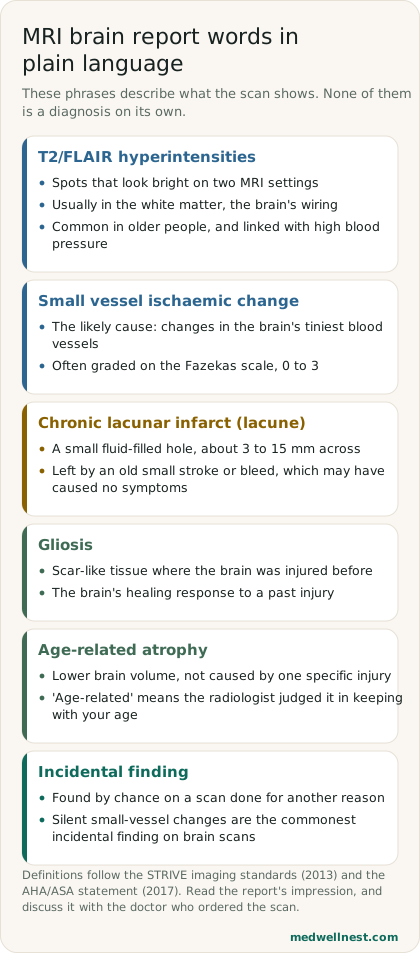
Many people meet these words on a scan done for something else, such as headaches, dizziness or a fall. Radiologists call that an incidental finding: something seen by chance, not the reason for the scan. The American Heart Association (AHA) and American Stroke Association describe silent disease of the small blood vessels as the most common incidental finding on brain scans, and as a common problem of ageing.
The numbers show how ordinary it is. A 2010 review in the BMJ found that white matter hyperintensities appear in 11% to 21% of adults around the age of 64, and in 94% of people at the age of 82. A few bright spots on a report of someone in their sixties or older is therefore a very common result, not a diagnosis. It is still worth understanding, because the same finding is linked with future stroke and memory problems across large groups of people.
02
T2/FLAIR hyperintensities and small vessel ischaemic change
T2 and FLAIR are two of the settings, or sequences, an MRI scanner uses. A hyperintensity is simply a spot that looks brighter than the tissue around it. The STRIVE imaging standards define a white matter hyperintensity of presumed vascular origin as a signal change in the white matter that is bright on T2-weighted images such as FLAIR, and that is not a fluid-filled hole.
"Small vessel ischaemic change" is the radiologist's view of the likely cause: wear in the brain's tiniest arteries, which supply the deep white matter. STRIVE notes that these spots are usually on both sides and fairly symmetrical, are common in older people, and are strongly linked with vascular risk factors. The same finding goes by many names; the STRIVE team counted 50 different terms in 940 papers, including leukoaraiosis, white matter changes and age-related white matter disease.
03
Fazekas grade 1, 2 or 3
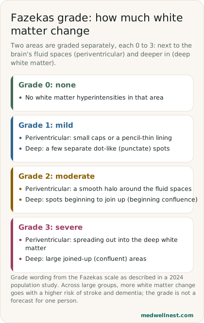
Many reports add a Fazekas grade, a simple visual score from 0 to 3. Two areas are often graded separately: the white matter next to the brain's fluid-filled spaces, called periventricular, and the deeper white matter. Grade 1 means small caps or a pencil-thin lining next to the fluid spaces, or a few separate dot-like spots deeper in. Grade 2 means a smooth halo, or spots beginning to join up. Grade 3 means changes spreading into the deep white matter, or large joined-up areas.
A higher grade means more white matter change, and across large groups more change goes with a higher risk of stroke and dementia. The grade is not a forecast for one person. The grade describes how far the change extends on the scan; it does not measure symptoms or memory.
04
Gliosis, atrophy and a chronic lacunar infarct
A lacune, often reported as a chronic lacunar infarct, is a different finding. STRIVE defines it as a round or oval fluid-filled hole deep in the brain, between 3 mm and about 15 mm across, left by an old small stroke or bleed in the territory of one tiny artery. Some of these small strokes happen without anyone noticing, which is why they are sometimes called silent. "Chronic" means old, not happening now.
Gliosis is the brain's scar-like reaction to an earlier injury: support cells called glia multiply around the damaged area. It marks where something happened in the past, such as a small stroke, an injury or inflammation. Age-related atrophy means the brain volume is a little lower than expected, without one specific injury to explain it; the words "age-related" mean the radiologist judged it in keeping with your age.
05
What the spots mean for stroke and memory
The 2010 BMJ review pooled long-term studies. People with white matter hyperintensities had about 3.3 times the risk of stroke, 1.9 times the risk of dementia and 2.0 times the risk of death compared with people without them. The AHA statement adds that white matter hyperintensities are linked with future stroke independently of other vascular risk factors.
The same review warns that these pooled figures should not be used to estimate the risk of one patient with white matter hyperintensities. The studies measured the spots in different ways, and the figures describe groups, not individuals. What the finding does is flag a brain whose small blood vessels deserve protection from now on.
06
What to do next: blood pressure first
Blood pressure matters most. A 2015 review by two small-vessel specialists calls high blood pressure the most important modifiable risk factor for stroke and the strongest known vascular risk factor for small vessel disease, and notes that multiple trials show treating high blood pressure prevents first strokes. The BMJ review advises that finding these spots should prompt a careful check of the risk factors for stroke and dementia.
In practice that means measuring your blood pressure properly, often with home readings, and checking blood sugar, cholesterol, weight and smoking. The AHA says no trials have yet tested treatments aimed specifically at silent small-vessel disease, but that ordinary stroke prevention is indicated in people who have it. Any medicine is a decision for your doctor; do not start one on your own because of a report.
An old report does not make new symptoms safe. If you notice a sudden change, use B.E. F.A.S.T. from the CDC: Balance loss, Eye or vision changes, Face drooping, Arm weakness, Speech trouble, Time to call emergency services. Note the time the symptoms began, because it guides treatment.
When to act
Emergency — act now
Sudden loss of balance, sudden vision trouble, a drooping face, weakness or numbness of an arm or leg on one side, slurred or strange speech, sudden confusion or a sudden severe headache: call emergency services now and note the time it started, whatever an earlier MRI report said.
Routine — see a doctor
A report mentioning white matter hyperintensities, small vessel ischaemic change, a Fazekas grade, gliosis or an old lacune, with no new symptoms: book a routine visit with the doctor who ordered the scan to review the report and check your blood pressure, blood sugar and cholesterol.
Values mentioned in this story
Each one opens a visual guide with its normal range and what moves it — upload a report and we place your own numbers on the same scale.
Sources
- The clinical importance of white matter hyperintensities on brain MRI: systematic review and meta-analysis — BMJ 2010pmc.ncbi.nlm.nih.gov
- Neuroimaging standards for research into small vessel disease (STRIVE) — Lancet Neurology 2013pmc.ncbi.nlm.nih.gov
- Prevention of Stroke in Patients With Silent Cerebrovascular Disease — AHA/ASA Scientific Statement 2017ahajournals.org
- Pharmacological treatment and prevention of cerebral small vessel disease — International Journal of Stroke 2015pmc.ncbi.nlm.nih.gov
- Fazekas visual assessment scales and white matter hyperintensities in a population study — PMCpmc.ncbi.nlm.nih.gov
- Stroke Signs and Symptoms — CDCcdc.gov
Share this story
Curious where your own values stand?
Two minutes from upload to a full visual picture. Free to analyze.