In short
A peripheral blood smear is a look at your blood cells under a microscope, often ordered when a CBC is abnormal. This guide puts phrases like microcytic hypochromic, anisocytosis, target cells and atypical lymphocytes into plain words and says which findings, such as blasts or schistocytes, need prompt follow-up.
Emergency if: Your smear or blood report mentions low platelets, schistocytes or blasts and you have bleeding that will not stop, a fever with a very low white cell count, or you feel very unwell: go to an emergency department now. The JMA review lists low platelets with bleeding, anaemia with low platelets and haemolysis, and fever with severe neutropenia as patterns needing urgent or immediate assessment.
On your report
MCV, RDW, Hemoglobin, Platelet Count +1 more
On this page
01
What is a peripheral smear test and why was it ordered?
MedlinePlus explains that a blood smear is a test that checks a sample of your blood under a microscope to see whether your blood cells look normal. A drop of blood is spread on a glass slide and treated with a stain, and a laboratory professional looks at the size, shape and number of the red cells, white cells and platelets. It is also called a peripheral smear or peripheral blood film, and computer programs may help. No special preparation is needed.
You may need one if a complete blood count (CBC) gave abnormal results, because a smear can confirm the result and show what the cells look like, including their colour. MedlinePlus also lists symptoms of a blood cell problem as reasons: fatigue, jaundice, nosebleeds or unusual bleeding, easy bruising, fever, bone pain and swollen lymph nodes. It is used to follow treatment for anaemia or blood cancer, and to look for parasites such as malaria in people who live in or travel to places where it is common.
A 2026 review explains the other reason: automated analysers do not reliably separate immature or abnormal cells, so a smear is done when the analyser raises a flag or the numbers do not fit the clinical picture. MedlinePlus is clear that a smear alone usually cannot diagnose a condition. Your doctor combines it with your history, symptoms and other tests, such as the CBC and the differential count.
02
What do microcytic, normocytic and macrocytic mean?
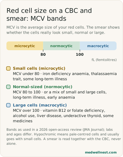
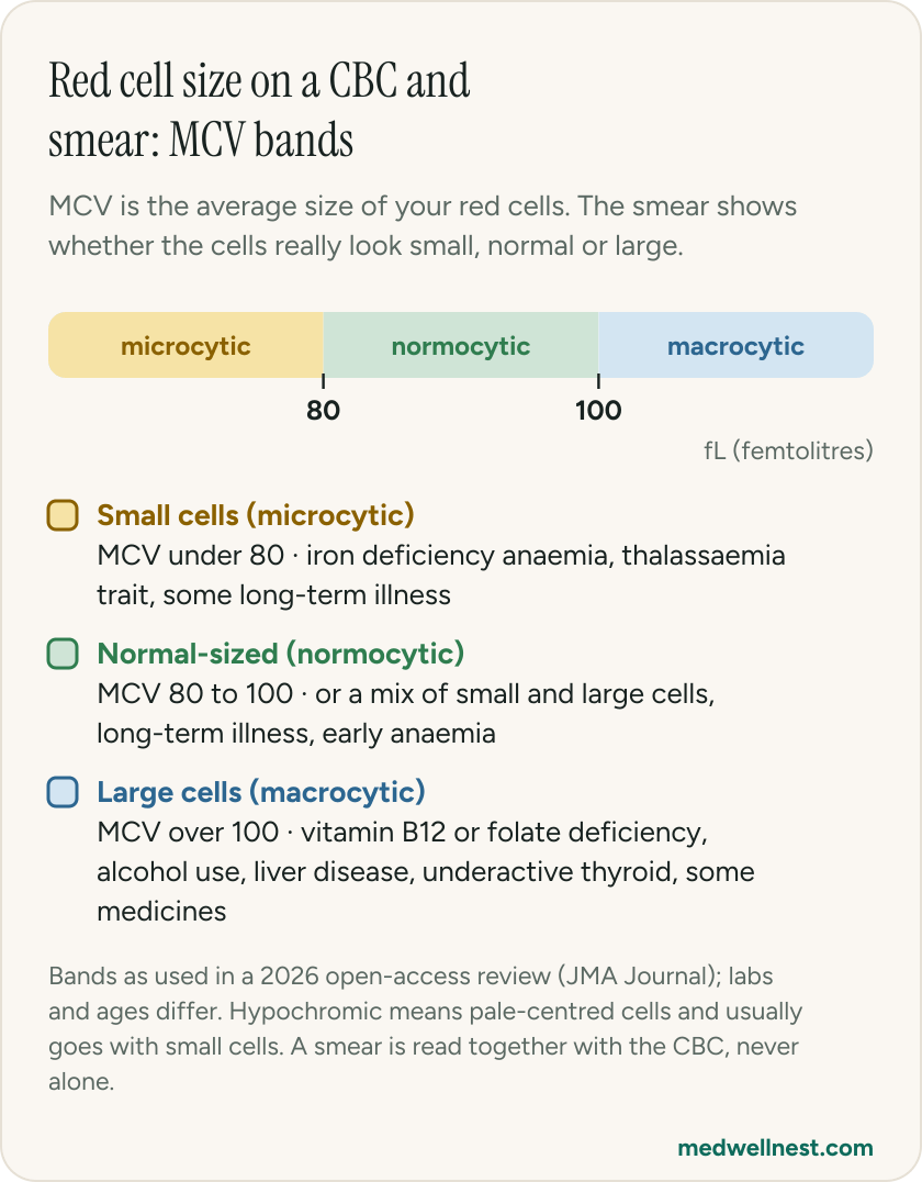
These words describe the size of the red cells. A 2026 review in the JMA Journal says the MCV gives a broad classification into microcytic, normocytic and macrocytic patterns, with low MCV under 80 fL, normal 80 to 100 fL and high over 100 fL. On the slide, a reviewer can also compare each red cell with a normal lymphocyte: a paediatric review says red cells larger than normal lymphocytes are called macrocytosis and smaller ones are called microcytosis.
The same JMA review pairs the MCV with the red cell distribution width (RDW), which shows how much the sizes vary. Low MCV with a high RDW fits iron deficiency anaemia, sideroblastic anaemia or combined causes, and low MCV with a normal RDW fits thalassaemia trait or some anaemia of long-term illness. A high MCV with a high RDW fits vitamin B12 or folate deficiency or myelodysplastic syndromes, and a high MCV with a normal RDW fits alcohol use, liver disease, an underactive thyroid or some medicines. A normal MCV does not exclude a problem if the RDW is high, because small and large cells can cancel out.
So a word like microcytic is a clue that leads to the next test, such as ferritin or haemoglobin analysis, not a diagnosis. Our stories on anaemia types and on ferritin show what usually comes next, and the one on the CBC explains MCV and RDW.
03
What are hypochromic, normochromic, anisocytosis and poikilocytosis?
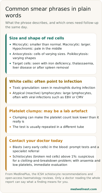
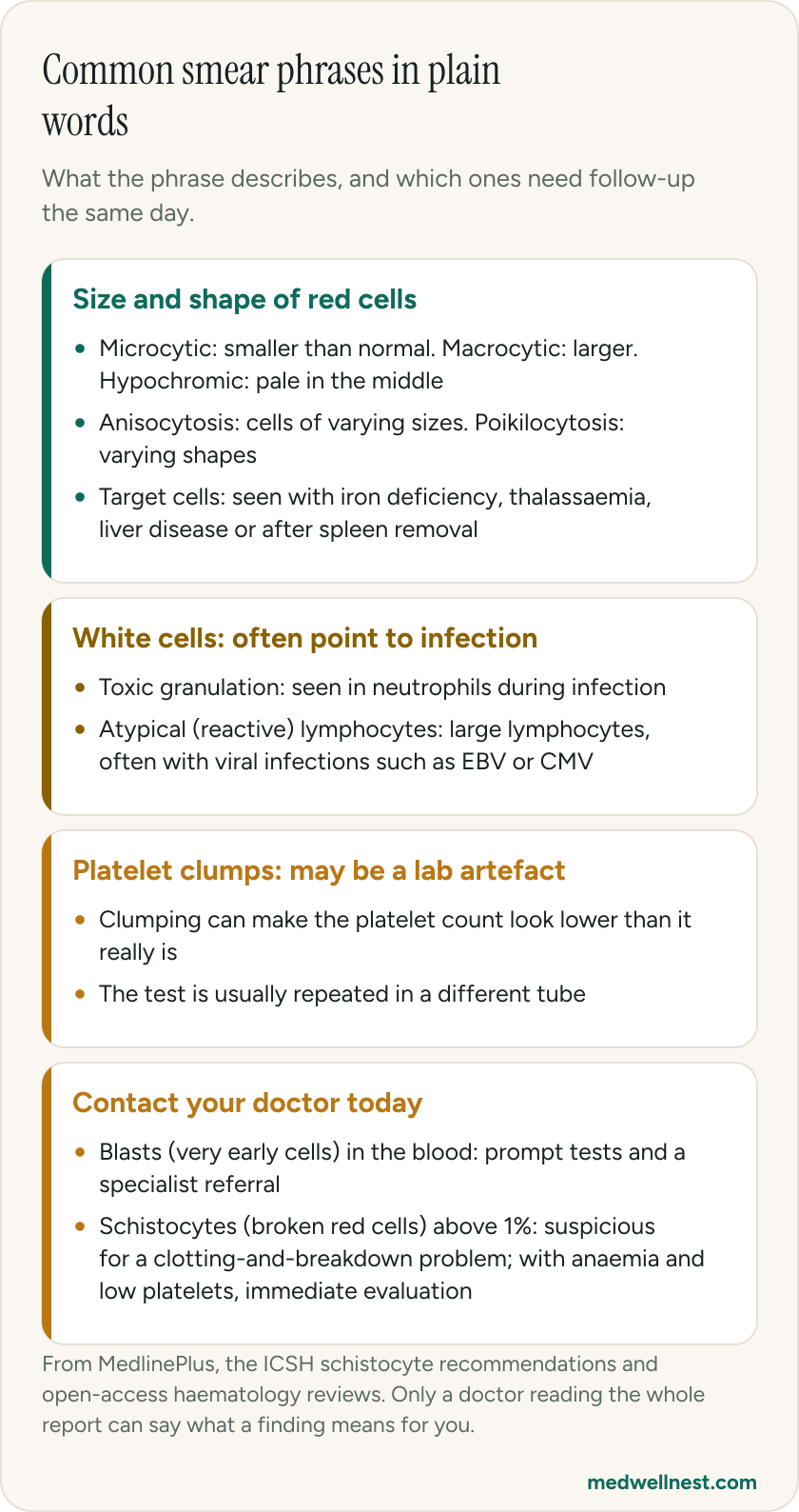
MedlinePlus says normal red cells are of similar size and colour and are lighter in the centre. Hypochromic means the cells are paler than usual. Hypochromia is seen in iron deficiency and thalassaemia. A paediatric review says microcytic red cells are mostly hypochromic. Normochromic simply means the colour is normal, and a report that says normocytic normochromic is describing cells of normal size and colour.
Anisocytosis means red cells of varying sizes, and poikilocytosis means red cells of varying shapes. The paediatric review links the RDW to the variation in cell diameter, which it calls anisocytosis. MedlinePlus adds that abnormalities may be graded on a 4-point scale: 1+ means one quarter of cells are affected, 2+ means half, 3+ means three quarters and 4+ means all of them. These words describe the slide. They do not say why the cells look this way.
04
What do target cells mean? Does it mean thalassaemia?
Not by itself. MedlinePlus lists the conditions that may go with target cells: abnormal haemoglobin (haemoglobinopathies), a deficiency of an enzyme called lecithin cholesterol acyltransferase, iron deficiency, liver disease and removal of the spleen. A paediatric review gives a similar list, with haemoglobin C, D and E, thalassaemia, iron deficiency anaemia and liver disease, and notes that target cells may indicate an illness or may be caused by artefacts.
So target cells plus small red cells makes the doctor think of thalassaemia or iron deficiency and ask for iron tests and often a haemoglobin analysis. Our stories on the HPLC report and on thalassaemia explain those next steps. Other named cells have their own clues: MedlinePlus links sphere-shaped cells to immune haemolytic anaemia or hereditary spherocytosis, and teardrop cells to severe iron deficiency, thalassaemia major or marrow scarring.
05
What do toxic granulation and atypical lymphocytes mean?
These are white cell findings, and they often point towards infection. A paediatric review says toxic granulation, vacuoles and Döhle bodies in neutrophils are smear findings that support infection. The report is not naming the germ: the same review says toxic granulation is typical of some bacterial infections, but a doctor still decides what is going on with your symptoms and other tests. Our story on the differential count explains neutrophils and lymphocytes.
Atypical lymphocytes are also called reactive lymphocytes. The paediatric review says they are generally much larger than normal lymphocytes, and that more than 10% of them on a smear supports a viral infection such as EBV or CMV, or toxoplasmosis. It also warns that inexperienced readers may confuse them with blasts. That is a reason the slide is read by a pathologist or haematologist and not decided from the analyser alone. A smear can also show malaria parasites, so tell your doctor if you have fever and have been in a malaria area.
06
My smear says platelet clumps. Are my platelets really low?
Maybe not. The JMA review says pseudothrombocytopenia, a falsely low platelet count, is most commonly caused by platelets clumping together in the test tube, including clumping that depends on the EDTA anticoagulant in the tube. It should be suspected when the count is unexpectedly low, the analyser flags clumps, or the result does not match how you are, especially if you have no bleeding symptoms. It can mimic severe thrombocytopenia and lead to unnecessary treatment or urgent referral.
The usual check is the smear itself and a repeat count in a tube with a different anticoagulant, such as citrate or heparin. A paediatric review says an unexpectedly low platelet count should always be confirmed on a smear using blood taken without EDTA. If the repeat count is truly low, our story on low platelet counts explains the causes beyond dengue. Whatever the smear says, report bleeding that will not stop or new, unusual bruising to a doctor without waiting.
07
Which smear findings need prompt follow-up: blasts and schistocytes?
Blasts are very early, immature blood cells. The JMA review calls circulating blasts, unexplained abnormal cells or persistent analyser flags a critical red flag that may indicate a haematological cancer and needs prompt diagnostic evaluation, including a smear and a specialist referral. A report word is still not a diagnosis. The paediatric review says early precursor cells on a smear may also be found in myelofibrosis, severe bleeding and haemolysis, and that reactive lymphocytes can be mistaken for blasts, so a specialist confirms the finding.
Schistocytes are fragments of red cells broken by mechanical damage in the circulation. The ICSH 2021 update gives 1% or less as the reference range for healthy adults and says more than 1% schistocytes on a film is suspicious for thrombotic microangiopathy (TMA), a condition of small clots and cell breakdown. MedlinePlus lists an artificial heart valve, thrombotic thrombocytopenic purpura, disseminated intravascular coagulation and haemolytic uraemic syndrome as causes. The JMA review says anaemia with low platelets and signs of haemolysis calls for immediate evaluation, that schistocytes on the smear support TMA, and that their absence does not rule TMA out.
If your report mentions blasts or schistocytes, do not wait for a routine appointment. Contact the doctor who ordered the test the same day, and go to an emergency department now if you are bleeding or very unwell.
When to act
Emergency — act now
Your smear or blood report mentions low platelets, schistocytes or blasts and you have bleeding that will not stop, a fever with a very low white cell count, or you feel very unwell: go to an emergency department now. The JMA review lists low platelets with bleeding, anaemia with low platelets and haemolysis, and fever with severe neutropenia as patterns needing urgent or immediate assessment.
Same-day — call promptly
Your report mentions blasts, schistocytes, or says a haematology opinion is advised: contact the doctor who ordered the test today. The JMA review says circulating blasts need prompt diagnostic evaluation and specialist referral, and the ICSH says schistocytes above 1% are suspicious for thrombotic microangiopathy. Do not wait for a routine appointment.
Routine — see a doctor
Your report has words such as microcytic, hypochromic, anisocytosis, target cells, toxic granulation or platelet clumps and you feel well: show it to the doctor who ordered it and ask what the CBC and smear together suggest, which test is next (for example ferritin, vitamin B12 or a repeat platelet count) and when to repeat the CBC.
Values mentioned in this story
Each one opens a visual guide with its normal range and what moves it — upload a report and we place your own numbers on the same scale.
Sources
- Blood Smear — MedlinePlus Medical Testmedlineplus.gov
- Blood smear — MedlinePlus Medical Encyclopediamedlineplus.gov
- How to Interpret the Complete Blood Count in Daily Practice — JMA Journal 2026pmc.ncbi.nlm.nih.gov
- What does a hemogram say to us? — paediatrics journal, 2020pmc.ncbi.nlm.nih.gov
- 2021 update of the ICSH recommendations for identification, diagnostic value, and quantitation of schistocytespubmed.ncbi.nlm.nih.gov
Share this story
Curious where your own values stand?
Two minutes from upload to a full visual picture. Free to analyze.