In short
Osteophytes, joint-space narrowing, sclerosis and a grade from 0 to 4: here is what a knee X-ray report is describing, why the grade often does not match the pain, and what actually helps a knee with osteoarthritis.
On your report
ESR, CRP, Rheumatoid Factor (RA Factor)
On this page
01
What the grade on your report is describing
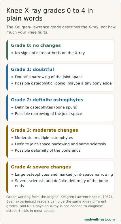
Many knee X-ray reports end with a Kellgren-Lawrence grade, a scale from 0 to 4 first published in 1957. It describes how the bones of the knee look on the film: whether there are bony outgrowths at the joint edges, whether the gap between the bones has narrowed, and whether the bone ends have changed shape. It is a description of the picture, not a measure of how much the knee hurts or how well it works.
Cartilage itself does not show up on an X-ray. What the X-ray shows is the space where cartilage sits between the thigh bone and the shin bone, so a narrower space is taken as a sign that cartilage has thinned. Grading is also less exact than it looks. In one study of the scale, agreement between even experienced graders was as low as 0.36, which the reviewers call poor reliability, so two readers can give the same knee different grades.
02
The report words, one at a time
An osteophyte is a bony outgrowth, often called a bone spur, at the edge of a joint. Joint-space narrowing means the gap between the bones is smaller than expected. Sclerosis, sometimes written as subchondral sclerosis, means the bone just under the cartilage looks denser and whiter than usual. Deformity of the bone ends means the joint surfaces have changed shape.
The grades build from these words. Grade 0 has no changes. Grade 1 is doubtful: possible narrowing and possibly a tiny bony edge. Grade 2 has definite osteophytes with possible narrowing. Grade 3 has moderate, multiple osteophytes, definite narrowing, some sclerosis and possible deformity. Grade 4 has large osteophytes, marked narrowing, severe sclerosis and definite deformity of the bone ends.
03
Why the grade and the pain often disagree
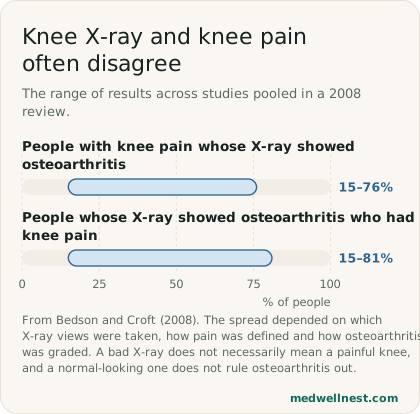
A 2008 review by Bedson and Croft pulled together studies that compared knee X-rays with knee pain. Among people with knee pain, the share whose X-ray showed osteoarthritis ranged from 15% to 76%. Among people whose X-ray showed osteoarthritis, the share who had knee pain ranged from 15% to 81%. The authors found that the results depended on which X-ray views were taken, how pain was defined and how osteoarthritis was graded.
In practice this means a grade 3 or 4 report does not predict a life of pain, and a grade 0 or 1 report does not mean your pain is imagined. The X-ray is one piece of information; how the knee feels and what you can do with it are what treatment aims to change.
04
Doctors usually do not need the X-ray to diagnose it
The NICE guideline on osteoarthritis (NG226) says to diagnose it clinically, without imaging, in people who are 45 or over, have joint pain related to activity, and have either no morning stiffness or morning stiffness lasting no longer than 30 minutes. It advises against routine imaging unless there are atypical features or features that suggest another diagnosis. The NHS similarly says X-rays or blood tests are not usually needed, but may be used to rule out other causes such as rheumatoid arthritis or a fracture.
NICE's committee found no evidence that imaging helps diagnose osteoarthritis and agreed it rarely adds anything useful for planning non-surgical treatment. It also advises against routine X-rays to follow the condition over time. The main place for imaging is when joint surgery is being planned or considered, to confirm how severe the structural changes are.
05
What actually helps
NICE names two core treatments: therapeutic exercise and, if you are living with overweight, weight loss, along with information and support. Exercise should be tailored to you, for example strengthening the muscles around the knee and general aerobic fitness, and supervised sessions can help. NICE warns that pain may increase when you first start, and explains that regular, consistent exercise is still good for the joint and that sticking with it over the long term brings more benefit.
On weight, NICE says that losing weight improves quality of life and physical function and reduces pain, that any amount of weight loss is likely to help, and that losing 10% of body weight is likely to be better than 5%. Medicines are there to support this, not replace it. NICE says there is no strong evidence of benefit for paracetamol, and that steroid injections into the joint give only short-term relief, of 2 to 10 weeks.
06
Red flags: when a painful knee may not be osteoarthritis
NICE lists features that call for a closer look, including a recent injury, morning stiffness that lasts a long time, symptoms or deformity that worsen quickly, a hot swollen joint, or anything that suggests infection or cancer. Long morning stiffness, the NHS notes, can be a sign of rheumatoid arthritis rather than osteoarthritis.
A hot, swollen joint needs particular care, because it can be septic arthritis, an infection inside the joint. The NHS describes sudden severe pain in a joint, being unable to move it or put weight on it, hot, swollen or red skin over it, and often a fever, although not everyone has one. It calls septic arthritis serious and says it should be treated as soon as possible; most people are admitted to hospital for treatment.
When to act
Same-day — call promptly
A joint that suddenly becomes very painful, hot, swollen or red, that you cannot move or stand on, especially with a fever or feeling unwell: get urgent medical help today. This can be septic arthritis, a joint infection that needs treatment as soon as possible. After an injury, a knee you cannot put weight on, that is badly swollen or has changed shape, or that locks or gives way: get it checked today.
Routine — see a doctor
Knee pain after a recent injury, morning stiffness that lasts well over 30 minutes, or pain or deformity that is getting worse quickly: see a doctor. These are the features for which NICE says imaging or other tests may be needed.
Osteoarthritis pain that is limiting your walking, work or sleep despite exercise: see your doctor or a physiotherapist about a structured exercise plan, weight support if needed, and other options.
Values mentioned in this story
Each one opens a visual guide with its normal range and what moves it — upload a report and we place your own numbers on the same scale.
Sources
- Osteoarthritis in over 16s: diagnosis and management (NG226) — NICEnice.org.uk
- The discordance between clinical and radiographic knee osteoarthritis — BMC Musculoskeletal Disorders, PMCpmc.ncbi.nlm.nih.gov
- Assessing radiographic knee osteoarthritis: an online training tutorial for the Kellgren-Lawrence grading scale — MedEdPORTAL, PMCpmc.ncbi.nlm.nih.gov
- Osteoarthritis — NHSnhs.uk
- Septic arthritis — NHSnhs.uk
Share this story
Curious where your own values stand?
Two minutes from upload to a full visual picture. Free to analyze.