In short
A grade 2 meniscus signal is not a tear, and many tears seen on MRI are not what causes the pain. Here is what meniscus grades, tear types, ACL sprain grades, bone bruise and effusion mean on a knee MRI, when surgery comes up, and which knee problems cannot wait.
Emergency if: A hot, red, swollen, painful knee with fever, or a knee you suddenly cannot move because of pain and swelling: go to an emergency department now. A joint infection is an emergency that can damage the joint.
On your report
CRP, ESR
On this page
01
What a knee MRI shows, and what it cannot tell you
An MRI of the knee shows the soft parts an X-ray cannot: the two menisci, which are C-shaped cushions of cartilage between the thigh bone and the shin bone, the ligaments such as the ACL, the smooth cartilage that covers the ends of the bones, and the bone marrow. The report describes what the radiologist sees. It does not say what is causing your pain, and that difference matters more than any single word on it.
Many findings turn up in knees that do not hurt. The BMJ's 2017 expert panel on knee arthroscopy wrote that meniscus tears are common, usually incidental findings, and unlikely to be the cause of knee pain, aching or stiffness. The Society of Skeletal Radiology (SSR), in a 2026 consensus on how to report the meniscus, calls signal inside the meniscus frequent in people without symptoms. So read your report together with your symptoms and your doctor's examination, not on its own.
02
Meniscus grades 1, 2 and 3: only grade 3 reaches the surface
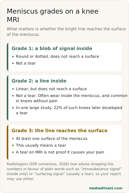
A healthy meniscus looks dark on MRI. Brighter areas inside it are called signal, and reports have long graded them. The SSR consensus gives the classic scheme: grade 1 is a globular or dotted signal that does not reach a surface of the meniscus; grade 2 is a linear signal that does not reach a surface; grade 3 extends to at least one surface. Only grade 3 is read as a tear: the consensus says signal that reaches the surface generally represents one.
Grade 2 is the one that worries people, and it is not a tear. The SSR says signal inside the meniscus commonly corresponds to mucoid degeneration or calcium deposits, a form of wear. In data from a large study of knees, the Osteoarthritis Initiative, a linear grade 2 signal carried a higher risk of later developing a tear, but most such knees did not go on to tear; 22% did.
Your report may not use numbers at all. The SSR now recommends that clinical reports drop numerical grading and describe the key features instead, because grading systems are often modified and cause confusion. Words such as intrasubstance signal, meaning inside only, or surfacing signal, meaning it reaches the surface, carry the same message.
03
Tear types: horizontal, radial, bucket-handle, complex
If the report does describe a tear, it usually names its pattern. The SSR lists the main patterns as horizontal, longitudinal-vertical, radial and complex. A radial tear runs across the width of the meniscus from its inner free edge towards the outer rim, and it is called complete or full-width when it reaches the rim. A complex tear has more than one tear plane. A bucket-handle tear is a long lengthwise tear, and the report may say its inner piece is displaced into the centre of the knee.
The pattern hints at the cause. The SSR notes that a horizontal tear in an older knee with worn cartilage may suggest a degenerative process, while a lengthwise tear in a young person after an injury may suggest a traumatic one, although many tears involve both. Words such as degenerative tear or maceration, which is used for worn meniscus tissue that has lost volume or frayed, point towards wear rather than a single injury. The report may also say which third of the meniscus is involved: the SSR describes a gradient from the outer rim, which has a blood supply, to the inner free edge, which does not.
04
ACL: sprain, partial tear or complete tear
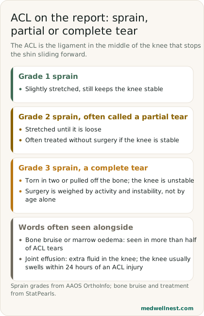
The anterior cruciate ligament (ACL) runs through the middle of the knee and stops the shin bone sliding forward. The American Academy of Orthopaedic Surgeons (AAOS) grades ligament injuries in three steps and says MRI is used to see how much damage there is. A grade 1 sprain is slightly stretched but still keeps the knee stable. A grade 2 sprain stretches the ligament until it is loose, and is often called a partial tear. A grade 3 sprain is a complete tear: the ligament is torn in half or pulled off the bone, and the knee is unstable.
StatPearls, a peer-reviewed medical reference, puts the accuracy of MRI for ACL injury at a sensitivity of 86% and a specificity of 95%, so the report is strong evidence but not the last word; your doctor also tests how the knee moves. Two other phrases often appear with an ACL injury. Bone bruise, or bone marrow oedema, is bruising inside the bone from the impact, and StatPearls says it is seen in more than half of ACL tears. Joint effusion means extra fluid in the knee; the AAOS says the knee usually swells within 24 hours of an ACL injury.
05
When surgery comes up, and when it usually does not
For wear-related knee problems in middle age, the evidence is against keyhole surgery. The BMJ panel made a strong recommendation against arthroscopy in nearly all patients with degenerative knee disease, which it defined as knee pain, particularly over 35, with or without a meniscus tear, arthritis on imaging, clicking or catching, or a sudden start. It cited a 2016 trial in which arthroscopy was no better than exercise therapy for a degenerative medial meniscus tear. Other options it lists include watchful waiting, weight loss if needed, physiotherapy-led exercise and pain relief.
The panel named one possible exception: a true locked knee, one that cannot be fully straightened. That needs an orthopaedic assessment rather than waiting it out.
An ACL tear is weighed differently. StatPearls says treatment without surgery suits partial tears and people who do not play sports that involve cutting or pivoting, while reconstruction is indicated for a complete tear in active, high-demand people at any age, including over 40. The AAOS says the decision also depends on how unstable the knee feels, how worn it already is and your activity level. No timetable for recovery fits everyone, so ask your own surgeon or physiotherapist what to expect.
06
Knee problems that cannot wait for a report
A hot, swollen, painful knee with fever can be septic arthritis, an infection inside the joint. StatPearls calls it an orthopaedic emergency that can damage the joint, and says the knee is the joint most often affected in adults. Doctors test fluid drawn from the joint; blood tests such as CRP and ESR support the diagnosis but cannot settle it on their own. Go to an emergency department straight away; do not wait for an MRI appointment.
MedlinePlus, the National Library of Medicine's health site, also advises contacting a doctor if you cannot put weight on the knee, the knee locks, buckles or will not straighten, it looks deformed, the pain is severe even at rest, or there is pain, swelling, numbness or a bluish colour in the calf below it. After an injury, the first-aid steps for sprains and suspected fractures are in our first-aid guide.
When to act
Emergency — act now
A hot, red, swollen, painful knee with fever, or a knee you suddenly cannot move because of pain and swelling: go to an emergency department now. A joint infection is an emergency that can damage the joint.
Same-day — call promptly
You cannot put weight on the leg after an injury, the knee is locked and will not straighten, it looks deformed, or the calf below it is painful, swollen or bluish: see a doctor today.
Routine — see a doctor
Your MRI shows a grade 2 signal, a degenerative tear or a partial ACL tear and the knee is stable: book a visit to go through the report and ask about a physiotherapy-led exercise plan before deciding on surgery.
Values mentioned in this story
Each one opens a visual guide with its normal range and what moves it — upload a report and we place your own numbers on the same scale.
Sources
- Standardized MRI nomenclature for knee meniscal lesions: SSR consensus panel report — Skeletal Radiology, 2026pmc.ncbi.nlm.nih.gov
- Arthroscopic surgery for degenerative knee arthritis and meniscal tears: a clinical practice guideline — BMJ Rapid Recommendations, 2017bmj.com
- Anterior Cruciate Ligament (ACL) Injuries — AAOS OrthoInfoorthoinfo.aaos.org
- Anterior Cruciate Ligament Knee Injury — StatPearlsncbi.nlm.nih.gov
- Septic Arthritis — StatPearlsncbi.nlm.nih.gov
- Knee pain — MedlinePlus Medical Encyclopediamedlineplus.gov
Share this story
Curious where your own values stand?
Two minutes from upload to a full visual picture. Free to analyze.