In short
A late-pregnancy growth scan reports the baby's estimated weight and its centile, the head-down or breech position, where the placenta lies, and the fluid around the baby. Here is what each line means, which numbers doctors act on, and what the scan is not used for.
Emergency if: Your baby is moving less than usual, you cannot feel it moving, or its pattern of movements has changed: call your hospital or maternity unit immediately, even at night, and do not wait until the next day. Do not use a home heartbeat monitor to reassure yourself.
On your report
Hemoglobin, Fasting Glucose, Post-Prandial Glucose (PPBS)
On this page
01
What a third-trimester growth scan is for
The International Society of Ultrasound in Obstetrics and Gynecology (ISUOG), whose 2024 guideline covers this scan, lists what it can assess: whether the baby is alive and well, its presentation (which way it is lying), its anatomy and growth, the amount of amniotic fluid, where the placenta is, and blood flow on Doppler. Doctors also order it for particular reasons, such as bleeding, the baby moving less, waters breaking early, or a bump that measures small or large.
ISUOG says the timing, anywhere between 32 and 36 weeks, should be decided by the pregnancy's risk level and local resources. It notes that in low-risk pregnancies, a small or large baby may be picked up more accurately at 36 weeks than at 32, while higher-risk pregnancies may be scanned earlier. So two women at the same hospital may have their scans at different weeks for good reasons.
The report is a snapshot. Most of its numbers only mean something next to your due date, your earlier scans and your doctor's plan, so read it as the start of a conversation rather than a verdict.
02
EFW and the four measurements: BPD, HC, AC and FL
Four measurements make up the biometry. BPD (biparietal diameter) is the width of the head, HC is the head circumference, AC is the abdominal circumference, and FL is the femur, or thigh-bone, length. ISUOG says all four can be measured routinely to assess the baby's size.
EFW, the estimated fetal weight, is not weighed. It is calculated from those measurements with a formula, and ISUOG recommends the Hadlock-3 formula, which uses HC, AC and FL, in most situations. Because it is a calculation from images, a small error in any one measurement carries into the weight.
The head can be harder to measure accurately late in pregnancy, ISUOG notes, because it sits deeper in the pelvis. That is one reason a single EFW should not be treated as the baby's exact weight, and why doctors look at the AC and the trend across scans as well.
03
Centiles, and why the due date should not move
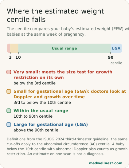
Beside each measurement, many reports print a centile, which compares your baby with babies at the same week of pregnancy. The 50th centile is the middle. ISUOG defines a baby as small for gestational age (SGA) when the EFW or AC is below the 10th centile, and large for gestational age (LGA) when it is above the 90th. A baby below the 3rd centile, or below the 10th with abnormal Doppler or slowing growth, meets its definition of fetal growth restriction (FGR).
SGA and growth restriction are not the same thing. Some small babies are simply small and healthy. Growth restriction means the baby is not reaching its potential, which is why doctors look at Doppler blood flow, the fluid, and repeat measurements rather than one number. Macrosomia, a very large baby at term, usually means a weight above a fixed line of 4000 or 4500 g.
Your due date (EDD) should come from your early scan. ISUOG says an 11 to 14 week measurement dates a pregnancy to within about 5 days, and that the pregnancy should not be re-dated using later scans. The gestational age a growth scan prints is the age the baby's size matches, so a baby measuring a week ahead or behind does not change your due date. Only if there was no earlier scan is a late scan used for dating, and then it can be out by about 15 days at 32 weeks.
04
Cephalic, breech, and where the placenta sits
Presentation tells you which part of the baby is lowest. Cephalic means head-down, which the NHS says most babies are by the time labour begins. Breech means bottom or feet first, and transverse means lying sideways. The NHS notes that many babies lie sideways earlier in pregnancy and turn head-down by themselves. If the baby is still breech at around 36 weeks, the NHS says you will usually be offered an external cephalic version (ECV), where a doctor tries to turn the baby by pressing on your abdomen, and your options for birth will be discussed.
Anterior, posterior and fundal describe which wall of the womb the placenta is attached to: the front, the back or the top. On its own, this simply records where it is. What doctors act on is how close the lower edge of the placenta is to the cervix, which ISUOG says every third-trimester scan should measure and document.
A placenta whose edge is less than 20 mm from the internal os (the inner opening of the cervix) is called low-lying, and one that covers the os is placenta praevia. If the edge is 20 mm or more away, ISUOG says vaginal birth is considered a safe option; if it is 10 to 20 mm away at 36 weeks, a vaginal birth may still be possible, and your doctor will plan the birth around it. If you have had a caesarean or other womb surgery and the placenta is low, ISUOG says it should be checked for placenta accreta, where it grows into the scar.
05
Placental grade, the fluid, and a cord around the neck
Some reports add a placental grade, 0 to 3, describing how calcified or mature the placenta looks. It is not among the things the ISUOG guideline lists for this scan to assess, and it is not one of the measurements used to judge growth. Treat it as a description rather than a result, and ask your doctor whether it changes anything for you before reading much into it.
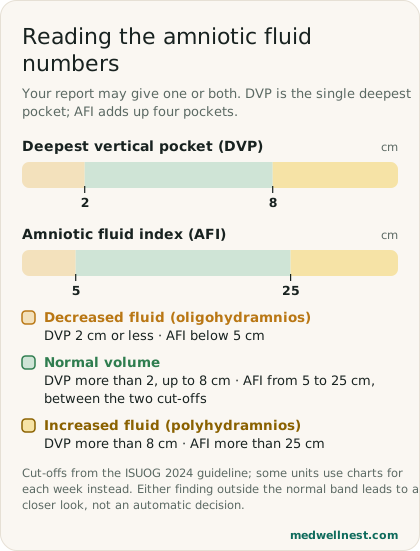
The fluid is reported as the deepest vertical pocket (DVP), the single deepest pool, or the amniotic fluid index (AFI), four pools added together. ISUOG counts a DVP of 2 cm or less, or an AFI below 5 cm, as decreased fluid, and a DVP above 8 cm, or an AFI above 25 cm, as increased. It prefers the DVP for calling fluid low, because using it led to fewer inductions with similar outcomes for babies.
A nuchal cord means the umbilical cord is looped around the baby's neck. Cleveland Clinic says it occurs in about 10% to 29% of pregnancies, rarely causes complications, and is often not even reported because it is so common. As long as blood flows well through the cord, which can be checked on Doppler, there is usually no concern, and at birth the loop can usually be slipped over the baby's head.
06
What the scan is not used for: the baby's sex
Nothing on a growth scan report tells you the sex of your baby, and nothing should. The Pre-Conception and Pre-Natal Diagnostic Techniques (PCPNDT) Act forbids anyone from using ultrasound to find out the sex of a fetus, and forbids anyone, including the person doing the scan, from telling the pregnant woman, her relatives or anyone else the sex by words, signs or any other means.
That includes hints dressed up as findings. Whether the placenta is anterior or posterior, and whether the heartbeat is fast or slow, are recorded for your baby's care, not as clues to the sex, and must not be used that way. A sonographer or doctor who signals the sex through them, or in any other way, is breaking the law. The scan exists to check that the baby is growing well and to plan a safe birth.
Bring the report to your next appointment with your questions written down: the EFW and AC centiles, the placental edge distance, the fluid measurement, and the presentation. Those are the lines your doctor will use to decide whether you need another scan, closer monitoring, or nothing more than your usual visits.
When to act
Emergency — act now
Your baby is moving less than usual, you cannot feel it moving, or its pattern of movements has changed: call your hospital or maternity unit immediately, even at night, and do not wait until the next day. Do not use a home heartbeat monitor to reassure yourself.
Heavy bleeding that soaks a pad soon after you put it on, or any bleeding with severe tummy pain, shoulder pain, or feeling faint or dizzy: get emergency care now.
Any bleeding at all if your scan showed a low-lying placenta or placenta praevia: go to your hospital or maternity unit now.
Same-day — call promptly
Light bleeding or spotting with no pain or only mild pain, when your scan did not show a low-lying placenta: contact your hospital or maternity unit the same day.
Routine — see a doctor
An EFW or AC below the 10th or above the 90th centile, fluid outside the normal range, a low-lying placenta, or a breech baby near 36 weeks: follow the plan your doctor sets, and ask when your next scan or check is due.
Values mentioned in this story
Each one opens a visual guide with its normal range and what moves it — upload a report and we place your own numbers on the same scale.
Sources
- ISUOG Practice Guidelines (2024): performance of third-trimester obstetric ultrasound scanisuog.org
- What happens if your baby is breech? — NHSnhs.uk
- Your baby's movements — NHSnhs.uk
- Vaginal bleeding in pregnancy — NHSnhs.uk
- Nuchal Cord — Cleveland Clinicmy.clevelandclinic.org
- The Pre-Conception and Pre-Natal Diagnostic Techniques (Prohibition of Sex Selection) Act, 1994indiacode.nic.in
Share this story
Curious where your own values stand?
Two minutes from upload to a full visual picture. Free to analyze.